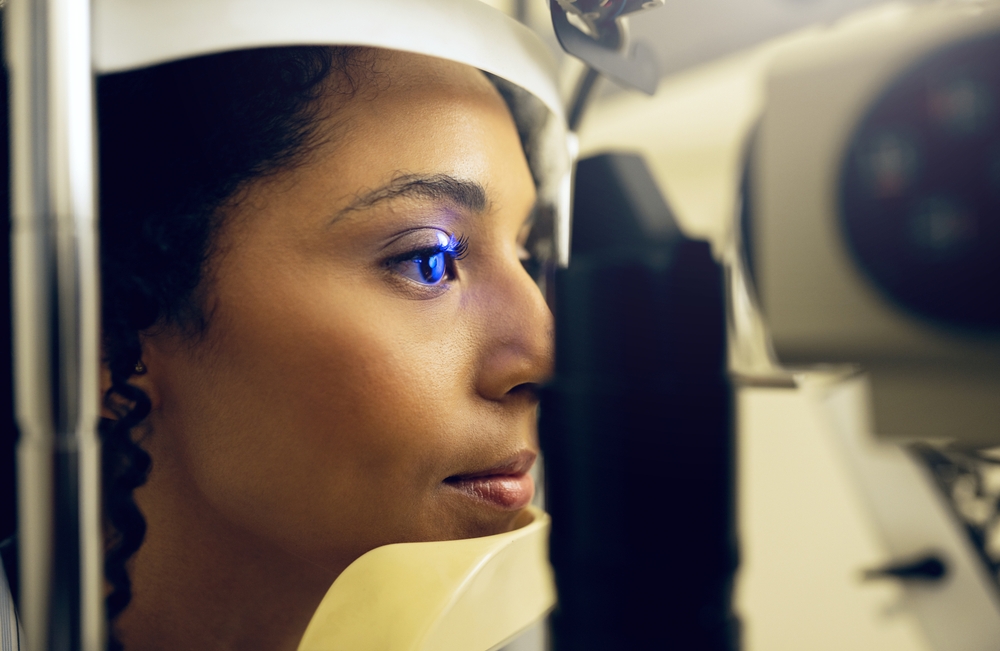
For individuals with diabetes, routine eye exams are crucial to maintain good eye health and prevent complications. Diabetes can lead to several eye-related issues, including diabetic retinopathy, glaucoma, and cataracts.
Why Regular Diabetic Eye Exams Matter
Diabetes-related eye conditions may not show symptoms in their early stages. Routine eye exams help catch changes before they progress, allowing for timely intervention that can preserve vision and prevent severe complications. Early diagnosis is key; whether it’s managing diabetic retinopathy, preventing glaucoma progression, or treating macular edema, catching these conditions early is the best way to protect your sight.
Visual Acuity Test
• What It Is: The visual acuity test is a standard part of most eye exams, assessing how clearly you can see at different distances.
• Why It’s Done: People with diabetes can experience changes in vision due to fluctuating blood sugar levels. Regular visual acuity testing helps track any gradual vision loss, which can be an early sign of diabetic retinopathy or other complications.
Dilated Eye Exam
• What It Is: During this test, eye drops are used to widen the pupils, allowing the optometrist to view the retina and optic nerve in detail.
• Why It’s Done: Diabetic retinopathy is a leading cause of vision loss in diabetics, and it affects the retina's blood vessels. A dilated eye exam allows the optometrist to detect any abnormal changes in these blood vessels, such as swelling, leakage, or new abnormal blood vessel growth. Early detection can prevent vision loss by enabling timely treatment.
Optical Coherence Tomography (OCT)
• What It Is: OCT is a non-invasive imaging test that provides detailed cross-sectional images of the retina.
• Why It’s Done: In diabetes, fluid can leak into the macula, the part of the retina responsible for sharp vision. OCT can detect swelling and fluid accumulation, known as diabetic macular edema, which can lead to vision impairment if not treated.
Fundus Photography
• What It Is: Fundus photography uses a specialized camera to capture images of the retina, optic nerve, and blood vessels.
• Why It’s Done: This test documents the retina's condition over time, allowing for monitoring of any progressive changes. In diabetic patients, it is particularly useful for detecting and tracking issues like diabetic retinopathy, glaucoma, and changes in the blood vessels.
Fluorescein Angiography
• What It Is: In this test, a fluorescent dye is injected into a vein in your arm, which travels to the blood vessels in your eye. Photos are taken as the dye circulates, highlighting any areas of leakage or abnormal blood vessels.
• Why It’s Done: Fluorescein angiography is helpful in diagnosing and monitoring advanced diabetic retinopathy and macular edema. By pinpointing areas of leakage, your optometrist can determine the best course of treatment, such as laser therapy or injections.
Tonometry
• What It Is: Tonometry measures the pressure inside the eye, known as intraocular pressure (IOP).
• Why It’s Done: People with diabetes are at a higher risk of developing glaucoma, a condition caused by increased eye pressure. Elevated IOP can damage the optic nerve over time, leading to vision loss if left untreated. Regular tonometry tests help in early detection and management of glaucoma.
Visual Field Test
• What It Is: This test measures your peripheral vision, assessing how much you can see out of the corner of your eyes.
• Why It’s Done: In diabetic patients, both diabetic retinopathy and glaucoma can affect peripheral vision. The visual field test can detect any loss in peripheral vision, which may indicate glaucoma or other retinal issues.
Schedule Your Eye Exam with Optimal Optometry Today
By understanding these essential tests and why they’re done, you can be proactive in your eye health. Regular check-ups are vital in managing your eye health and safeguarding your vision for years to come.
If you’re due for an eye exam or have noticed any changes in your vision, schedule your diabetic eye exam with Optimal Optometry. Visit our office in Ontario, California, or call (909) 563-3120 to book an appointment today.


